Ultrasound
Question 1. What is an Ultrasound?
Ultrasound is safe and painless, and produces pictures of the inside of the body using sound waves. Ultrasound imaging, also called ultrasound scanning or sonography, involves the use of a small transducer (probe) and ultrasound gel placed directly on the skin. High-frequency sound waves are transmitted from the probe through the gel into the body. The transducer collects the sounds that bounce back and a computer then uses those sound waves to create an image.
Ultrasound examinations do not use ionizing radiation (as used in x-rays), thus there is no radiation exposure to the patient. Because ultrasound images are captured in real-time, they can show the structure and movement of the body's internal organs, as well as blood flowing through blood vessels.
Ultrasound imaging is a noninvasive medical test that helps physicians diagnose and treat medical conditions.
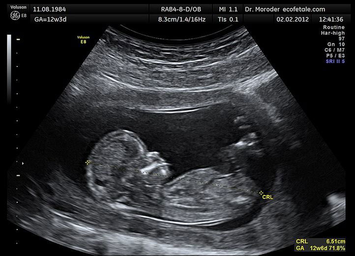
Question 2. What does the equipment look like?
diagram
Ultrasound scanners consist of a console containing a computer and electronics, a video display screen and a transducer that is used to do the scanning. The transducer is a small hand-held device that resembles a microphone, attached to the scanner by a cord. Some exams may use different transducers (with different capabilities) during a single exam. The transducer sends out high-frequency sound waves at 3-7 MHz (that the human ear cannot hear) into the body and then listens for the returning echoes from the tissues in the body. The principles are similar to sonar used by boats and submarines.
The ultrasound image is immediately visible on a video display screen that looks like a computer or television monitor. The image is created based on the amplitude (loudness), frequency (pitch) and time it takes for the ultrasound signal to return from the area within the patient that is being examined to the transducer (the device placed on the patient's skin to send and receive the returning sound waves), as well as the type of body structure and composition of body tissue through which the sound travels. A small amount of gel is put on the skin to allow the sound waves to travel from the transducer to the examined area within the body and then back again. Ultrasound is an excellent modality for some areas of the body while other areas, especially air-filled lungs, are poorly suited for ultrasound.
Question 3. How does the procedure work?
Ultrasound imaging is based on the same principles involved in the sonar used by bats, ships and fishermen. When a sound wave strikes an object, it bounces back, or echoes. By measuring these echo waves, it is possible to determine how far away the object is as well as the object's size, shape and consistency (whether the object is solid or filled with fluid).
In medicine, ultrasound is used to detect changes in appearance, size or contour of organs, tissues, and vessels or to detect abnormal masses, such as tumors.
In an ultrasound examination, a transducer both sends the sound waves into the body and receives the echoing waves. When the transducer is pressed against the skin, it directs small pulses of inaudible, high-frequency sound waves into the body. As the sound waves bounce off internal organs, fluids and tissues, the sensitive receiver in the transducer records tiny changes in the sound's pitch and direction. These signature waves are instantly measured and displayed by a computer, which in turn creates a real-time picture on the monitor. One or more frames of the moving pictures are typically captured as still images. Short video loops of the images may also be saved.
Question 4. How is the procedure performed?
For most ultrasound exams, you will be positioned lying face-up on an examination table that can be tilted or moved. Patients may be turned to either side to improve the quality of the images.
After you are positioned on the examination table, the radiologist (a physician specifically trained to supervise and interpret radiology examinations) or sonographer will apply a warm water-based gel to the area of the body being studied. The gel will help the transducer make secure contact with the body and eliminate air pockets between the transducer and the skin that can block the sound waves from passing into your body. The transducer is placed on the body and moved back and forth over the area of interest until the desired images are captured.
There is usually no discomfort from pressure as the transducer is pressed against the area being examined. However, if scanning is performed over an area of tenderness, you may feel pressure or minor pain from the transducer.
Once the imaging is complete, the clear ultrasound gel will be wiped off your skin. Any portions that are not wiped off will dry quickly. The ultrasound gel does not usually stain or discolour clothing.
Question 5. Who does the ultrasound procedure?
In PNG, the procedure is usually done by trained ultra-sonographers or radiologists. Occasionally, general doctors or specialist physicians and surgeons would perform the tests. If you are in doubt of your test results always have it done and interpreted by specialist radiologist.
Question 6. Who interprets the results and how do I get them?
A radiologist, a physician specifically trained to supervise and interpret radiology examinations, will analyze the images and send a signed report to your primary care physician, or to the physician or other healthcare provider who requested the exam. Usually, the referring physician or health care provider will share the results with you. In some cases, the radiologist may discuss results with you at the conclusion of your examination.
Follow-up examinations may be necessary. Your doctor will explain the exact reason why another exam is requested. Sometimes a follow-up exam is done because a potential abnormality needs further evaluation with additional views or a special imaging technique. A follow-up examination may also be necessary so that any change in a known abnormality can be monitored over time. Follow-up examinations are sometimes the best way to see if treatment is working or if a finding is stable or changed over time.
Question 7. What are the benefits vs. risks?
Benefits
Most ultrasound scanning is noninvasive (no needles or injections).
Occasionally, an ultrasound exam may be temporarily uncomfortable, but it should not be painful.
Ultrasound is widely available, easy-to-use and less expensive than other imaging methods.
Ultrasound imaging is extremely safe and does not use any ionizing radiation.
Ultrasound scanning gives a clear picture of soft tissues that do not show up well on x-ray images.
Ultrasound provides real-time imaging, making it a good tool for guiding minimally invasive procedures such as needle biopsies and fluid aspiration.
Risks
For standard diagnostic ultrasound, there are no known harmful effects on humans and no radiation like x-rays and CT scan.